
3D Bioprinter
We help life scientists push the frontiers of biofabrication
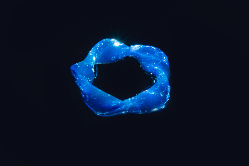
Leverages our contactless tomographic illumination technology to shape sensitive cells and biomaterials into biological systems, without impairing their viability. Volumetric printing not only preserves cells but also makes research more efficient by simplifying design iterations and statistical studies.

About The Technology

Rapid Volumetric Printing
Shaped light beams cure the full 3D volume simultaneously, allowing biological constructs to form in just a few seconds with exceptional speed and efficiency.

Gentle on Living Cells
Since the process is entirely light-based, it avoids the shear stress often associated with extrusion methods, helping preserve cell viability during fabrication.
About the Technology
Tomographic 3D Printing for Rapid,Cell-Friendly Biofabrication
Tomographic 3D printing rapidly solidifies photosensitive inks in three dimensions, using shaped light beams from multiple angles. As the entire build volume is illuminated simultaneously, centimeter-scale biological systems are produced in just a few seconds. After printing, the object is simply separated from the uncured ink and collected.
Our printing method is light-based, so it does not induce any shear stress on the printed cells. The remarkably low photoinitiator content (eg 1mg/mL LAP) and low light dose (<600 mJ/cm²) make tomographic bioprinting a cell-friendly technique.
.jpg)
Low-Stress, High-Compatibility Printing
The printing method uses remarkably low photoinitiator content, such as 1 mg/mL LAP, along with a low light dose below 600 mJ/cm², making tomographic bioprinting highly cell-friendly.
100%
Fast Fabrication
100%
Cell-Friendly Process
100%
Low Light Exposure
What It Enables
High-Speed Bioprinting for Complex Biological Structures
This technology enables the fast production of centimeter-scale biological systems with minimal cellular stress. By combining volumetric illumination with biocompatible printing conditions, tomographic bioprinting offers a promising solution for advanced tissue engineering, regenerative medicine, and next-generation biofabrication research.
Features
Specification
Specification | Unit |
|---|---|
Pixel size | 14µm |
Magnification before vial | 2x |
Indicative print time | 30s – 120s (depends on material) |
Build diameter | up to 6.3mm (standard) or 12.5mm (performance) |
Build height | ≥ 25mm |
Light intensity | 1 to 20mW/cm² (average at container) |
Wavelength | 405nm ± 5 nm (standard), 400nm ± 1 nm (performance)
Other wavelengths upon request |
Maximum rotation speed | > 60°/s |
Container materials | Autoclavable and reusable sealed glass containers |
Compatible materials | hydrogels, acrylics and silicones |
External footprint | 27cm x 30cm x 67cm |
Initial accessories kit | Precision chuck adaptor for vials
Vial extraction tool |
Example


Achieved Results
Title | Concentration | Viability | Construct size | Print time |
|---|---|---|---|---|
Human hepatic organoids | 5.10^6 cells/ml | > 95% after 10 days | Ø 6 × h 17 mm | 15.5s |
Human embryonic kidney cells (HEK 293) | 4.10^6 cells/ml | -- | Ø 8.1 × h 9 mm | 36s |
Mouse myoblasts (C2C12) | 10^6 cells/ml | > 90% after 7 days | Ø 7 × h 15 mm | 10 – 11s |
Normal human dermal fibroblasts (NHDF) | 10^6 cells/ml | > 90% after 7 days | 13 × 6.0 × 2.6 mm | 11.4s |
Equine mesenchymal stromal cells (MSCs) | 10^6 cells/ml | -- | Ø 8.5 × h 9.3 mm | 12.5s |
Equine articular chondroprogenitor cells (ACPCs) | 10^7 cells/ml | > 85% after 7 days | Ø 5.0 × h 1.0 mm | -- |